
The physical examination of the musculoskeletal system is essential for assessing posture, detecting deformities, and evaluating joint mobility. It ensures early diagnosis and effective treatment planning.
1.1 Overview of the Musculoskeletal System
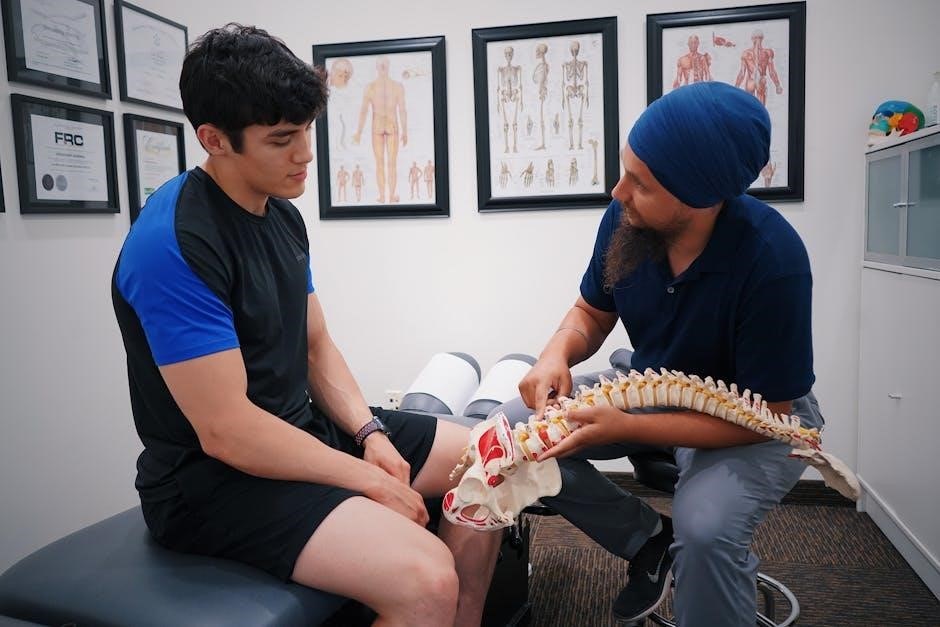
The musculoskeletal system comprises bones, muscles, joints, and ligaments, enabling movement, posture, and stability. It is divided into the axial and appendicular systems, working together to provide structural support and facilitate mobility. Bones act as the framework, while muscles generate force for movement. Joints allow articulation, and ligaments provide stability. This system is vital for daily activities and is often assessed through physical examinations to diagnose conditions affecting its components.
1.2 Importance of Physical Examination
Physical examination is crucial for diagnosing musculoskeletal disorders, guiding treatment, and monitoring recovery. It allows healthcare providers to identify abnormalities, assess function, and develop personalized care plans. Early detection of issues like deformities or tenderness can prevent complications. The exam also helps differentiate between various conditions, ensuring accurate diagnoses. Regular assessments improve patient outcomes and enhance the effectiveness of therapeutic interventions, making it a cornerstone of musculoskeletal healthcare.
Inspection and Observation
Inspection and observation involve assessing posture, alignment, deformities, and swelling. These visual examinations provide critical insights into joint mobility and overall musculoskeletal function, aiding in early diagnosis and treatment planning.
2.1 Posture and Alignment
Posture and alignment assessment involves evaluating the patient’s stance, spinal curvature, and symmetry. Proper alignment ensures optimal musculoskeletal function, while deviations may indicate underlying issues. Observing the head, shoulders, hips, knees, and ankles provides insights into structural integrity. Abnormalities in posture, such as kyphosis or scoliosis, can lead to discomfort or dysfunction. Accurate documentation of these findings aids in diagnosing conditions like uneven muscle development or joint misalignment, guiding targeted treatment strategies for improved patient outcomes and mobility.
2.2 Deformities and Swelling
Deformities and swelling are key indicators of musculoskeletal abnormalities. Inspection involves identifying visible abnormalities, such as fractures, joint dislocations, or congenital conditions. Swelling may result from inflammation, trauma, or fluid accumulation. Palpation helps confirm the presence of masses or effusions. Documenting the location, size, and symmetry of these findings aids in diagnosis. Such observations are critical for identifying conditions like fractures, arthritis, or soft tissue injuries, guiding further diagnostic steps and treatment interventions effectively.
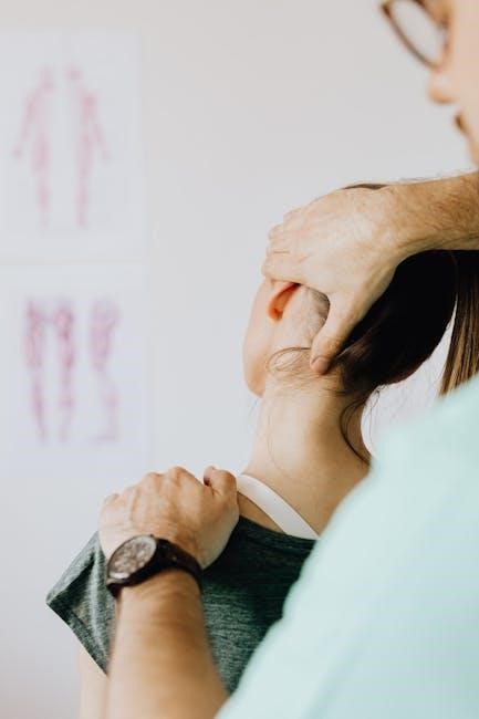
Palpation
Palpation involves manual assessment of soft tissues, muscles, and joints to detect tenderness, swelling, or abnormalities; It aids in identifying muscle atrophy, hypertrophy, and areas of pain or masses.
3.1 Muscle Atrophy and Hypertrophy
Muscle atrophy refers to a reduction in muscle mass, often due to disuse or neurological conditions. Hypertrophy is an increase in muscle size, typically from exercise. During palpation, atrophy may feel soft and diminished, while hypertrophy presents as firm and enlarged. Both conditions are assessed through comparative examination of symmetric muscle groups. Accurate identification aids in diagnosing underlying causes, such as nerve damage or overuse, guiding appropriate treatment strategies for musculoskeletal health.
3.2 Areas of Tenderness and Masses
Palpation identifies areas of tenderness, which may indicate inflammation or injury. Masses, such as tumors or cysts, are assessed for size, consistency, and mobility. Gentle pressure helps determine pain severity, while comparing affected and unaffected areas ensures accuracy. Documentation of these findings aids in differential diagnosis and treatment planning, crucial for addressing musculoskeletal disorders effectively and preventing complications.
Range of Motion Assessment
Range of motion assessment evaluates joint mobility, detecting limitations or abnormalities. It uses techniques like goniometers to measure movement, aiding in diagnosis and treatment planning effectively.
4.1 Upper Extremities
The range of motion assessment for upper extremities focuses on evaluating the shoulders, elbows, wrists, and hands. Active and passive movements are measured to identify limitations or abnormalities. Techniques such as flexion, extension, abduction, and rotation are commonly used. Goniometers or visual estimation may be employed to quantify movement. Abnormal findings, such as reduced mobility, may indicate conditions like arthritis or injury. Accurate measurements are crucial for diagnosing impairments and developing targeted rehabilitation plans to restore function and improve quality of life.
4.2 Lower Extremities
The range of motion assessment for lower extremities involves evaluating the hips, knees, and ankles. Techniques include flexion, extension, abduction, and rotation. Pain or limited mobility may indicate conditions like arthritis or injury. Goniometers are often used to measure joint angles accurately. Proper documentation of findings aids in diagnosing impairments and guiding rehabilitation. Early detection of issues in lower extremities is crucial for maintaining mobility and preventing further complications, ensuring effective treatment and improved patient outcomes.
Special Tests
Special tests in musculoskeletal examination help diagnose joint stability, neurological function, and soft tissue integrity. They are crucial for identifying specific injuries or conditions effectively.
5.1 Joint Stability Tests
Joint stability tests assess the integrity of ligaments and joint capsules. Common tests include the Lachman and Anterior Drawer tests for knee instability. These evaluations help identify laxity or tears, guiding treatment plans. They are essential for diagnosing conditions like ACL injuries, ensuring accurate assessments and targeted therapies. Proper technique is critical to avoid false positives and ensure patient safety during these procedures.
5.2 Neurological Examination
A neurological examination is crucial in assessing nerve function related to the musculoskeletal system. It involves testing reflexes, sensation, and muscle strength to identify nerve involvement. Techniques like the Hoffmann and Tinel’s signs help detect nerve compression or damage. This assessment aids in diagnosing conditions such as radiculopathy or neuropathy, ensuring comprehensive evaluation and appropriate treatment planning for patients with musculoskeletal and neurological symptoms.

Radiological and Diagnostic Techniques
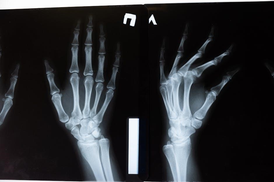
Radiological and diagnostic techniques, such as X-rays and MRIs, provide detailed insights into musculoskeletal structures, essential for accurate diagnosis and treatment planning.
6.1 Role of Radiomics in Musculoskeletal Assessment
Radiomics, a technique extracting quantitative features from medical images, enhances musculoskeletal assessment by analyzing X-rays and MRIs. Initially applied in oncology, it now aids in diagnosing musculoskeletal conditions, improving accuracy and enabling early detection of subtle changes. This non-invasive method provides objective, reproducible data, making it invaluable for monitoring progression and treatment response. Its integration into clinical practice offers a promising future for precise diagnosis and personalized treatment planning.
6.2 Imaging Modalities for Detailed Analysis
Imaging modalities like X-rays, MRIs, CT scans, and ultrasounds provide detailed insights into musculoskeletal structures. X-rays detect fractures and bone deformities, while MRIs offer soft tissue visualization, such as ligaments and tendons. CT scans combine bone and soft tissue imaging, ideal for complex injuries. Ultrasound is portable and effective for dynamic joint assessments. These tools complement physical exams, aiding in precise diagnosis and treatment planning by revealing internal abnormalities not visible through palpation or inspection alone.
Rehabilitation and Treatment Methods
Rehabilitation and treatment methods focus on restoring function and reducing pain through specially targeted exercises, physical therapy, and orthotic devices, enhancing recovery and improving quality of life.
7.1 Exercise Therapy
Exercise therapy plays a vital role in rehabilitation by improving strength, flexibility, and range of motion. Tailored programs address specific injuries, enhancing recovery and preventing future issues effectively.
7.2 Physical Therapy Modalities
Physical therapy modalities include heat, cold, ultrasound, and electrical stimulation, aiding in pain relief and tissue repair. These methods complement exercise therapy, enhancing recovery and restoring function.
The physical examination is crucial for early diagnosis and effective treatment. Future advancements in imaging and rehabilitation will enhance musculoskeletal care, significantly improving patient outcomes.
8.1 Summary of Key Findings
The physical examination of the musculoskeletal system is vital for detecting posture abnormalities, deformities, and joint mobility issues. It helps identify areas of tenderness and masses, enabling early diagnosis. Radiomics has emerged as a valuable tool for extracting detailed image features, aiding in precise assessments. Rehabilitation methods, including exercise therapy and physical therapy modalities, play a crucial role in restoring functionality and reducing pain. These findings underscore the importance of integrating clinical examination with advanced imaging for optimal musculoskeletal care and improved patient outcomes.
8.2 Future Directions in Musculoskeletal Examination
Future advancements in musculoskeletal examination may involve integrating advanced radiomics and AI-driven imaging tools for precise diagnostics. Wearable technology could enhance real-time monitoring of joint health and movement patterns. Personalized rehabilitation programs, tailored to individual needs, are expected to improve outcomes. Additionally, the development of non-invasive diagnostic methods and regenerative therapies could revolutionize treatment approaches, offering faster recovery and reduced pain management challenges in musculoskeletal care.
Leave a Reply
You must be logged in to post a comment.